Osteoblasts and osteoclasts
Want to learn more about the bone as an endocrine organ? Watch this video to learn about osteoblasts and osteoclasts, which produce and respond to hormone signals.
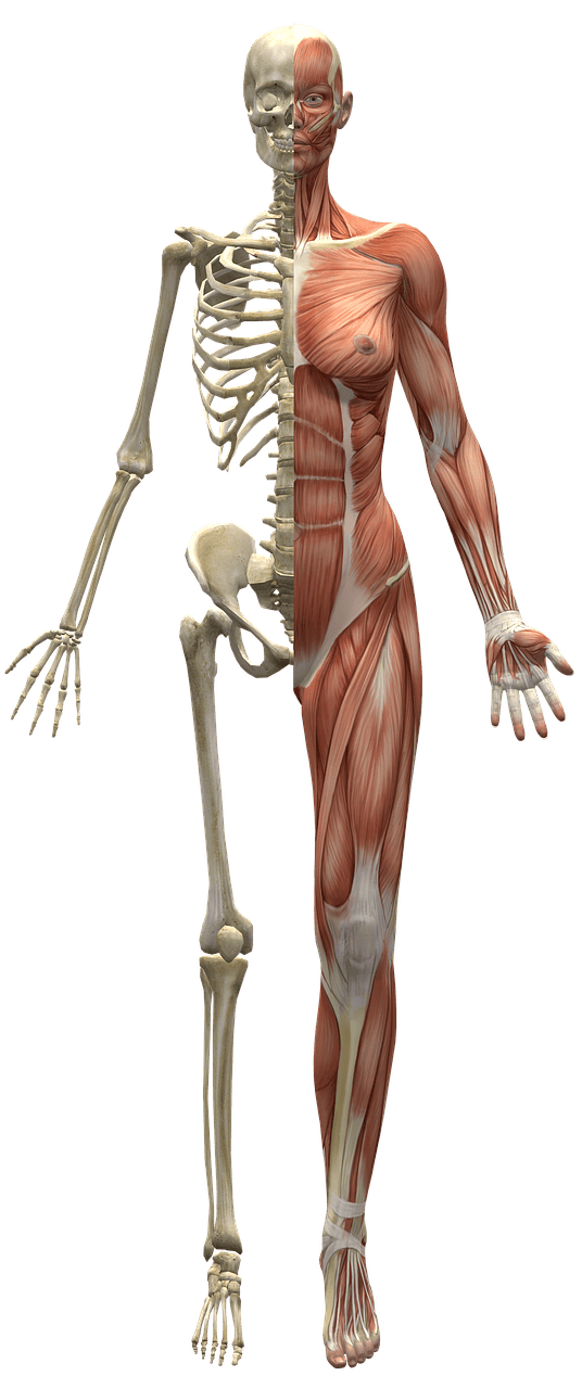
The skeletal system gives our bodies shape and support. Learning how it works helps us understand how our bodies move and stay strong. Use this resource to explore parts of the skeletal system and what they do. Also learn about a culturally responsive movement-based program to improve health outcomes, including skeletal health, for Aboriginal and Torres Strait Islander children.
The musculoskeletal system or locomotor system includes the bones, muscles, tendons and ligaments in the body. They work together to:
It consists of the skeletal and muscular systems. Here, we will look at the skeletal system. The next page will discuss the muscular system.
The skeletal system consists of all the bones in the body and its supporting tissues, including bones, cartilage, ligaments and joints. Together, these form the skeleton. The skeleton gives the body support and shape, and protects the internal organs.
Bones are strong structures that act as the framework for the skeleton. They play a crucial role in protecting the internal organs.
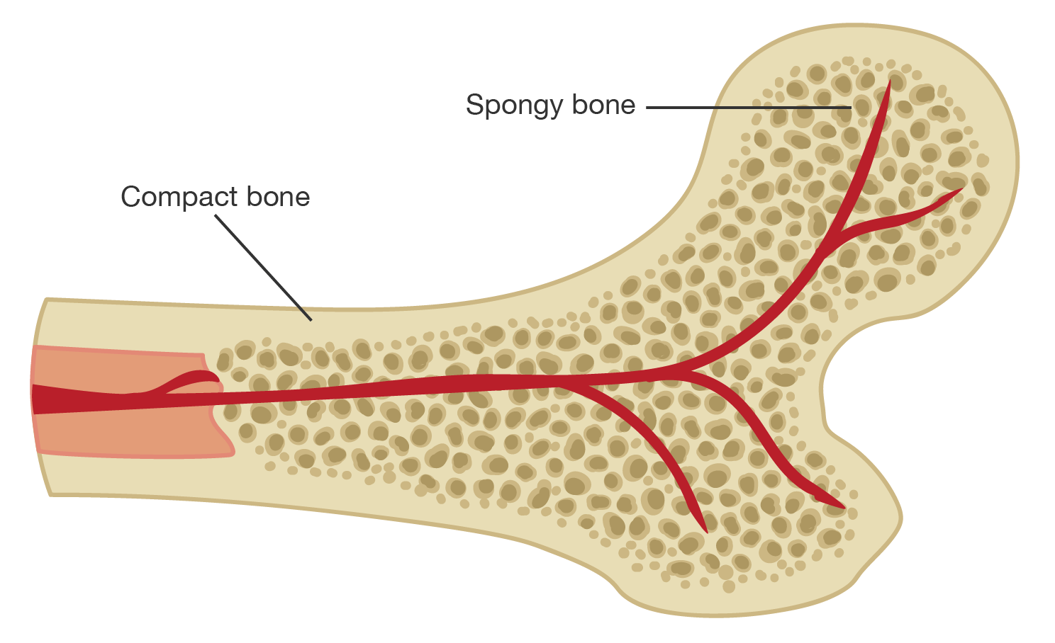
Bones have a dense outer layer called compact bone (also known as cortical bone) and a lighter, porous inner layer called spongy bone (also known as cancellous or trebacular bone). Both compact and spongy bone are mostly made of a mineral called hypoxyapatite, which is a form of calcium phosphate. This makes up around \(70\%\) of bone; the remaining \(30\%\) is a mix of cells and water.
A diagram of the end of a bone. Where the bone has been cut, there is red bone marrow. Extending through the core of the bone are blood vessels. The outer layer of bone is labelled "Compact bone" which is dense. The inner area of bone is labelled "Spongy bone", which is porous, as shown by the tiny holes throughout the structure.
Inside the bone
The spaces in the spongy bone are where bone marrow is found. Red marrow makes blood cells. Yellow marrow stores fat.
Blood vessels run through the compact and spongy bone, playing an important role in bone development, repair and maintenance.
Bone can be considered an endocrine organ because it produces and responds to hormones that regulate biochemical processes in the body.
The outer surface of the bone is covered in a thin membrane called the periosteum (“peri” meaning “around” and “osteon” meaning “bone”). It has blood vessels and nerves that help the bone grow and heal.
The overall structure of bone gives it strength, but also flexibility and the ability to absorb shock.
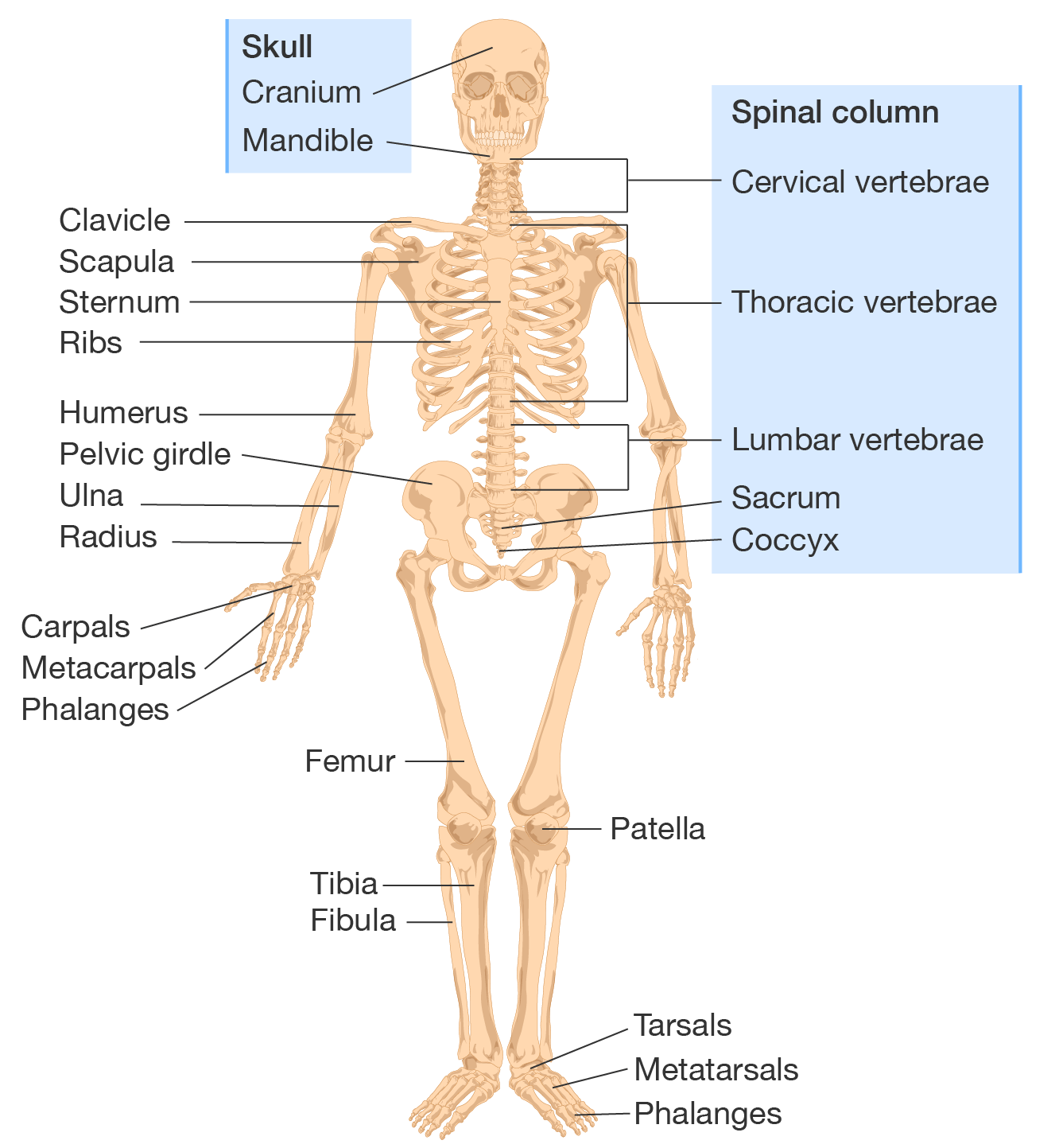
The image shows some of the key bones in the skeletal system.
A diagram of a labelled skeleton.
Bones in the skeletal system
Cartilage is a flexible type of tissue that cushions the joints and reduces friction between bones so you can move easily. You typically find cartilage covering the surfaces of bones at your joints. It also covers structures like the nose, ears and parts of the rib cage.
Cartilage is made of cells called chondrocytes (“chrondro” meaning “cartilage” and “cyte” meaning “cells”). These produce a gel-like substance that is both strong and flexible.
Unlike bone, the cartilage does not contain blood vessels. It gets nutrients from surrounding tissues. This means that when cartilage is damaged, it takes a lot longer to heal.
There are three main types of cartilage:
Bones are connected at joints by strong bands of tissue called ligaments. They provide stability by holding the bones together and allowing you to move in a controlled way, while also stopping you from making overextended or abnormal movements that could injure you.
Ligaments are made up of dense, fibrous connective tissue. This gives them strength and flexibility. They support movement and keep your joints aligned.
The joints connect bones to let us move. They are complex structures containing various tissue types. Joints include cartilage and ligaments.
We can classify joints based on their structure and the material that holds the bones together. There are three primary categories:
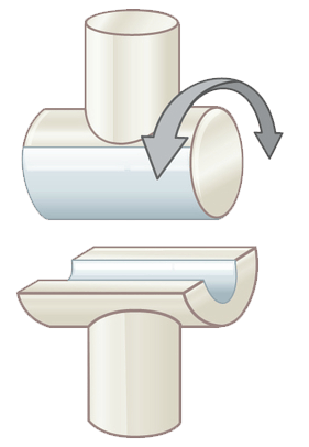
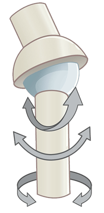
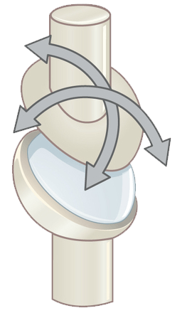
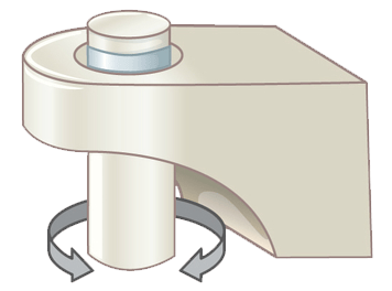
Synovial joints are the most common and familiar joints in the body. There are different types of synovial joints, which we have summarised in the following table.
| Type | Description | Movement | Example |
|---|---|---|---|
| Hinge | One bone with an end that rounds outwards fits into a bone with an end that rounds inwards | Open and close in one direction to allow bending and stretching
|
Knee |
| Ball-and-socket | One bone with a rounded end fits into the indentation of another bone | Can rotate and turn in almost any direction
|
Shoulder |
| Condyloid | One bone with an oval shape end fits into the indentation of another | Can flex, extend and move side to side, but not rotate
|
Wrist |
| Pivot | One bone with a round end fits into a ring-shaped tendon or bone | Can rotate around an axis
|
Neck |
| Planar | Two bones that are flat or slightly curved that slide over each other | Can slide or glide, but not rotate
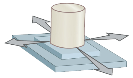
|
Carpal bones in the wrist |
| Saddle | One bone with an end that rounds outwards fits perpendicarly into a bone with an end that rounds inwards | Can move in any direction, but cannot twist or rotate
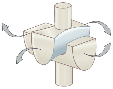
|
Thumb |
Joint images by OpenStax, licensed under CC BY 4.0.
Not all joints in your body can move. There are fibrous joints in your skull called 'sutures', which are not mobile... thank goodness!
A diagram of the skull showing the joints holding together the plates.
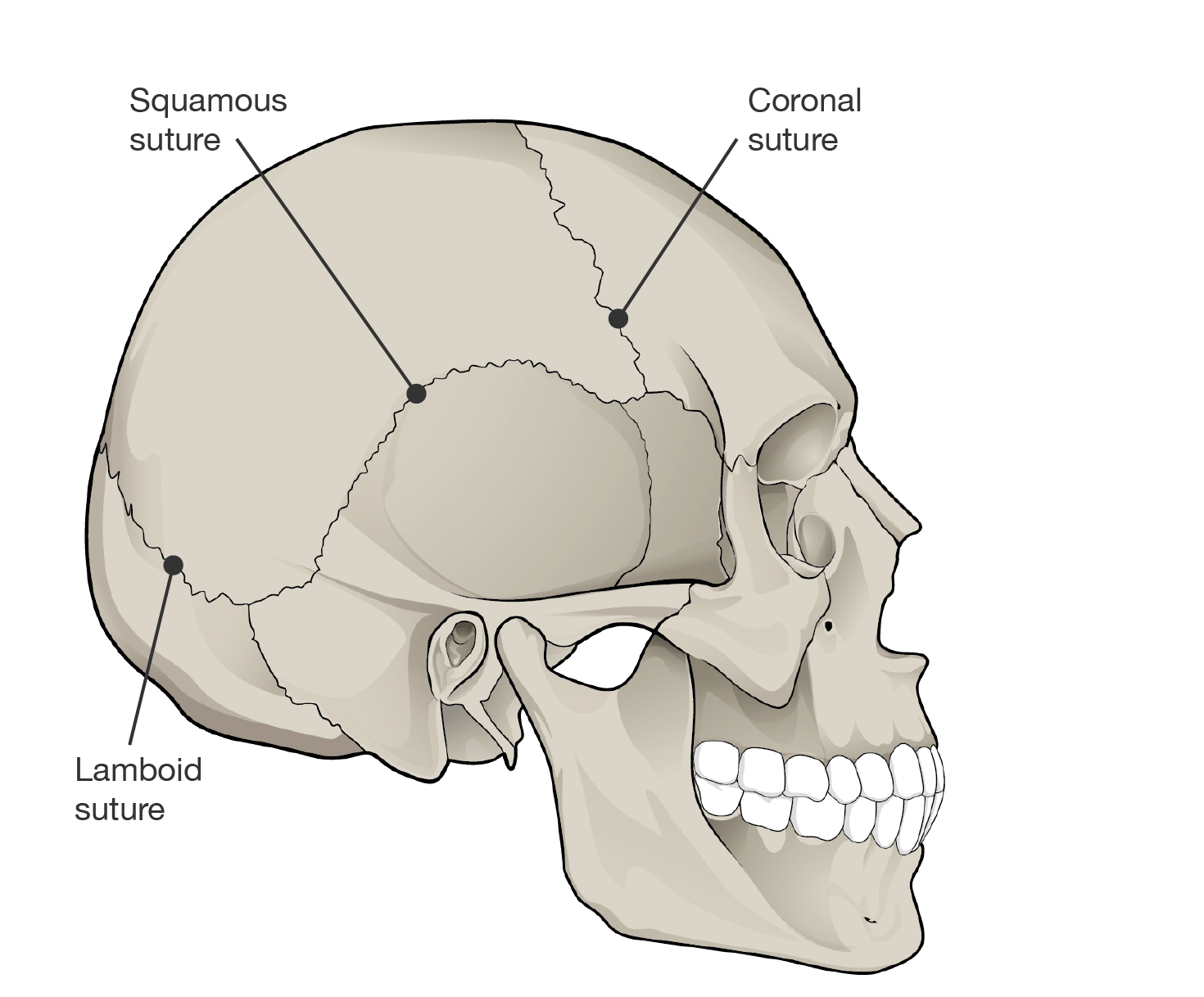
Joints in the skull
Indigenous knowledges in life science
Dr Kai Wheeler, a Ngarabal person and exercise scientist, has co-developed movement-based programs such as the 'Move2Smile with Culture' program—in partnership with the Yarrabah Aboriginal and Torres Strait Islander community—for Aboriginal and Torres Strait Islander children and adolescents. These programs embed Aboriginal and Torres Strait Islander ways of Knowing, Being and Doing, and focus on increasing participation in sport and physical activity, which is crucial for maintaining skeletal health.
By encouraging regular movement, Kai's research aims to reduce the risk of poor physical health outcomes that disproportionally affect young Indigenous people. This work not only enhances our understanding of how exercise benefits general health, including sleeping patterns and mental health, but also directly supports skeletal development in Aboriginal and Torres Strait Islander young people.
The components of the skeletal system and their functions are outlined in the table.
| Component | Function |
|---|---|
| Bones | Give structure to the body and protect the internal organs |
| Cartilage | Cushions joints and reduces friction between bones |
| Ligaments | Hold bones together |
| Joints | Allow the body to move |
See how well you understand the components and function of the skeletal system with a quick quiz.
Read the scenario and use the information provided to answer the questions in the quiz.
Dr. Pom Pompurin, a nutrition researcher, conducted a six-month study to examine the impact of calcium intake on bone health. He tracked the average weekly calcium intake of six healthy adults and assessed their bone health scores at the beginning and end of the study.
The bone health score, measured on a scale from 0 to 100, reflects changes in bone density and strength. The data is shown in the table.
| Average weekly calcium intake (g) | Bone health score (start) | Bone health score (end) |
|---|---|---|
| \(4.2\) | \(65\) | \(68\) |
| \(5.6\) | \(70\) | \(69\) |
| \(7.0\) | \(75\) | \(78\) |
| \(8.4\) | \(80\) | \(82\) |
| \(6.3\) | \(72\) | \(76\) |
| \(10.5\) | \(85\) | \(84\) |
Images on this page by RMIT, licensed under CC BY-NC 4.0
Osteoblasts and osteoclasts
Want to learn more about the bone as an endocrine organ? Watch this video to learn about osteoblasts and osteoclasts, which produce and respond to hormone signals.
The skeletal system
Explore a 3D model of the skeletal system using this interactive.
Learn the skeletal system game
Want to test your understanding of which bone goes where? Play this game!

RMIT University acknowledges the people of the Woi wurrung and Boon wurrung language groups of the eastern Kulin Nation on whose unceded lands we conduct the business of the University. RMIT University respectfully acknowledges their Ancestors and Elders, past and present. RMIT also acknowledges the Traditional Custodians and their Ancestors of the lands and waters across Australia where we conduct our business - Artwork 'Sentient' by Hollie Johnson, Gunaikurnai and Monero Ngarigo.
More information