National Geographic: Heart 101
Want to learn more about the heart and see how it works? Watch this video.
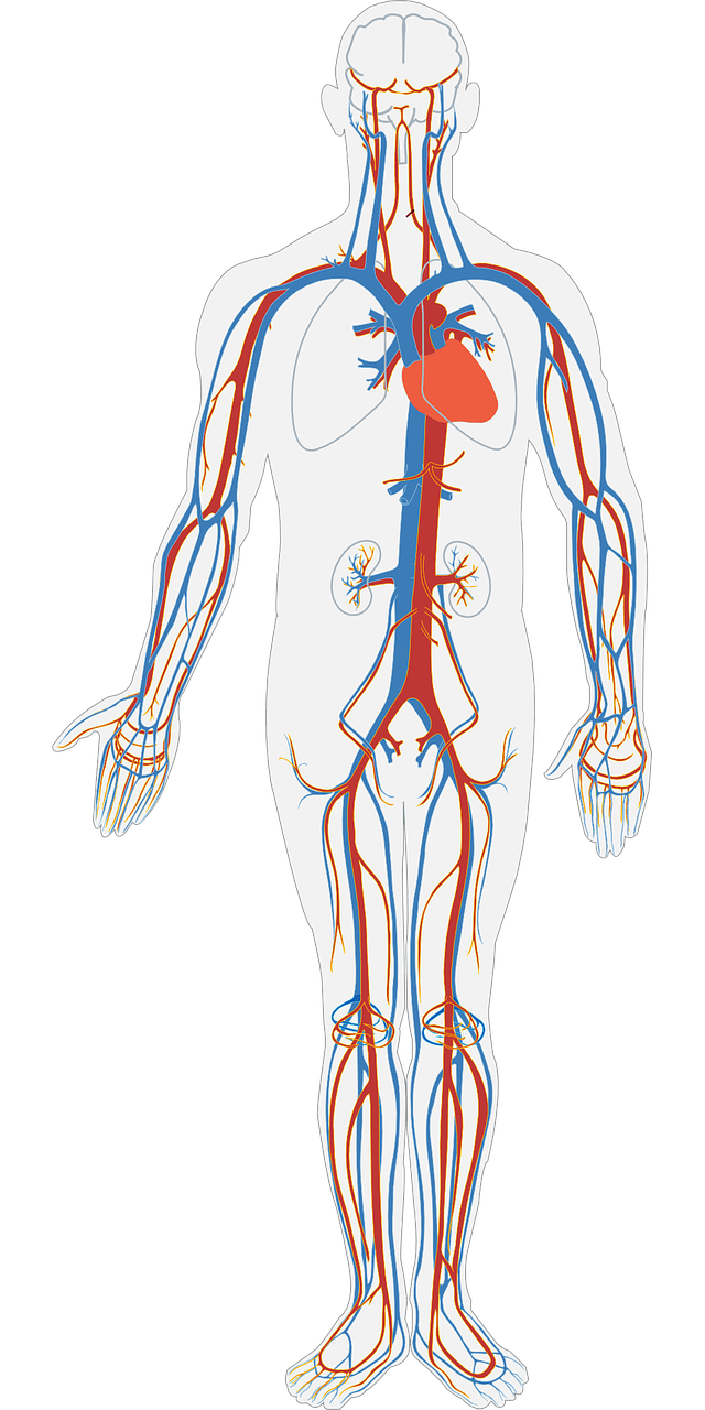
The circulatory system transports blood, oxygen and nutrients around the body. Understanding how it works helps us see how the body maintains its energy levels, responds to changes and stays healthy. Use this resource to explore the components and functions of the circulatory system.
The circulatory system or cardiovascular system consists of the heart, blood vessels and blood. They work together to sustain life by delivering vital substances to cells and removing waste products.
Blood is the fluid in our circulatory system. It is responsible for transporting oxygen, nutrients and hormones around the body. Its composition is summarised in the table.
| Component | Proportion |
|---|---|
| Red blood cells | \(44\%\) |
| White blood cells | \(<1\%\) |
| Platelets | \(<1\%\) |
| Plasma | \(55\%\) |
Click to reveal more detail about each component.
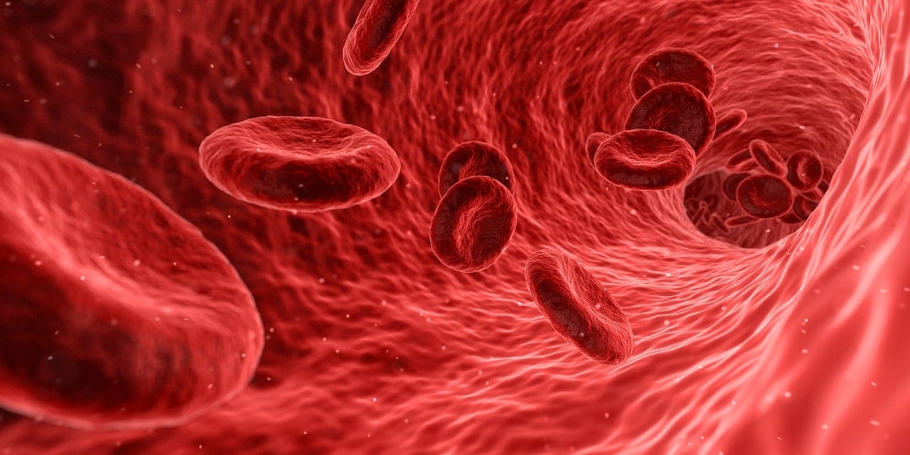
These cells contain a protein called haemoglobin, which contains iron that binds to oxygen. The presence of iron gives RBCs their colour.
RBCs have a biconcave shape where they curve inwards on both sides. This increases their surface area for gas exchange and allows them to move easily through blood vessels.
You can learn more about the immune response by going to Immunology.
When an injury occurs, they gather at the site and stick together to form a sort of plug that prevents excessive bleeding. They also release chemicals that activate during the clotting process to stabilise this plug. This gives the body some time to repair the tissue.
Plasma helps maintain blood pressure, regulate body temperature and balance pH levels to make sure that our cells function properly.
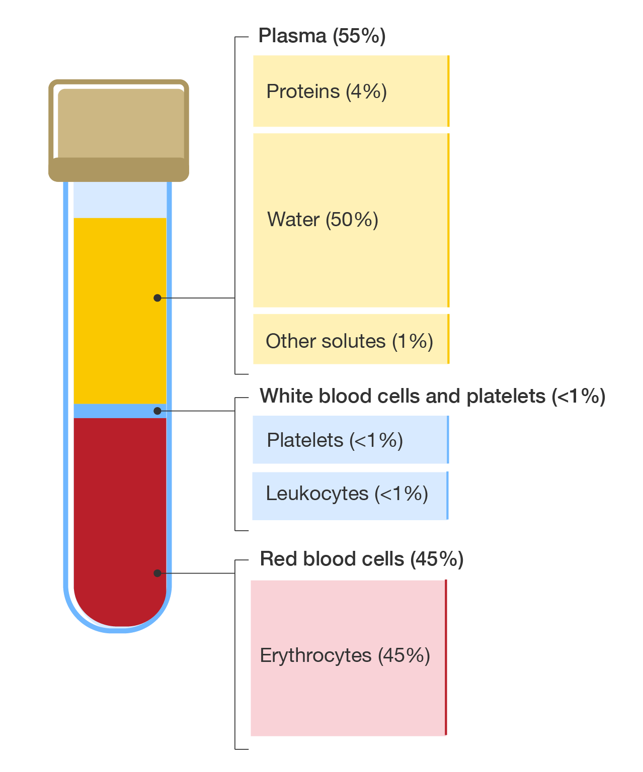
A diagram of a blood sample tube with the composition of blood shown.
The blood vessels are the network of pathways that carry blood throughout the body. There are three main types.
In the figure shown, arteries branch into smaller arterioles, which then branch into capillaries. The arteries and veins join at the capillary bed, then transition into venules, which then join up to form veins.
A diagram of the blood vessels showing how they are structured and connected.
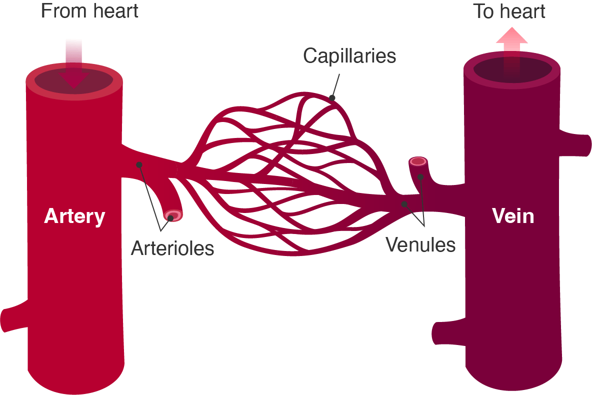
Blood vessels
Together, these vessels ensure that blood flows efficiently, delivering vital substances and removing waste from the body.
The heart is a muscular organ that pumps blood throughout the body to distribute oxygen, nutrients and hormones.
Animated Human Heart (inside) by raito via Sketchfab, licensed under Standard Sketchfab License
It has four chambers:
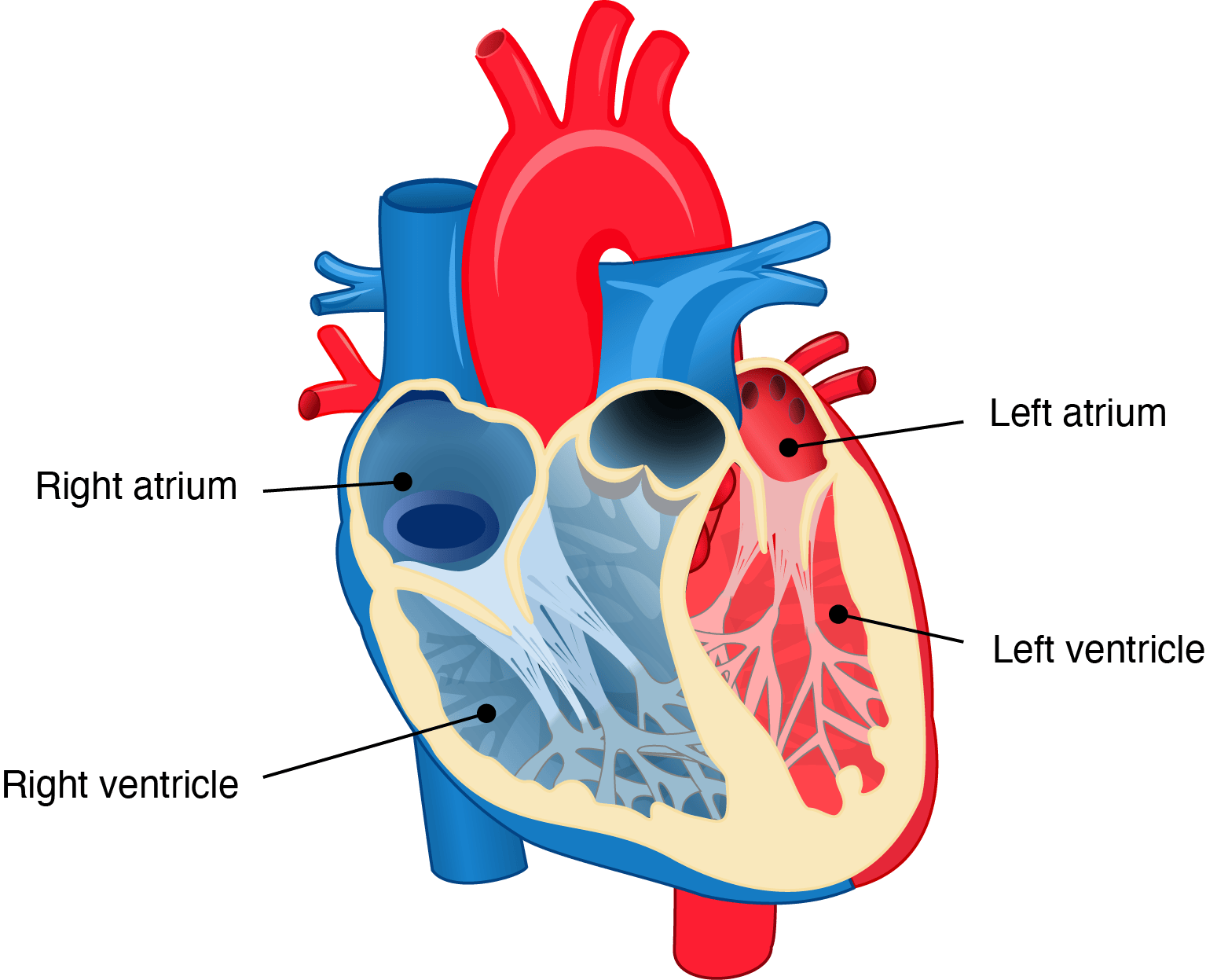
A diagram of the heart with the chambers visible.
The heart works in a coordinated way, with the right side of the heart involved in the pulmonary circulation and the left side involved in the systemic circulation.
A diagram showing the pathway of blood circulation. Blue pathways leading to the heart show deoxygenated blood and red pathways leading from the heart show oxygenated blood. The capillary beds are shown in purple to indicate gas exchange. The "Pulmonary circulation" involves the lungs, heart and pulmonary artery and vein. There is an arrow from the heart through the pulmonary artery to the lungs, then back to the heart via the pulmonary vein. The "Systemic circulation" involves the other major organs in the body. Arrows show movement of blood from the heart through the aorta to the capillary beds in the upper body, then back into the heart through the vena cava. Blood from the aorta also goes to the liver through the hepatic artery, then returns to the heart through the hepatic vein. Blood goes to the stomach and intestines, which gets filtered into the liver through the hepatic portal vein. The kidneys receives blood from the renal artery, which is returned to the heart via the renal vein. The lower body also has arteries and veins that transport blood to and from the lower body.
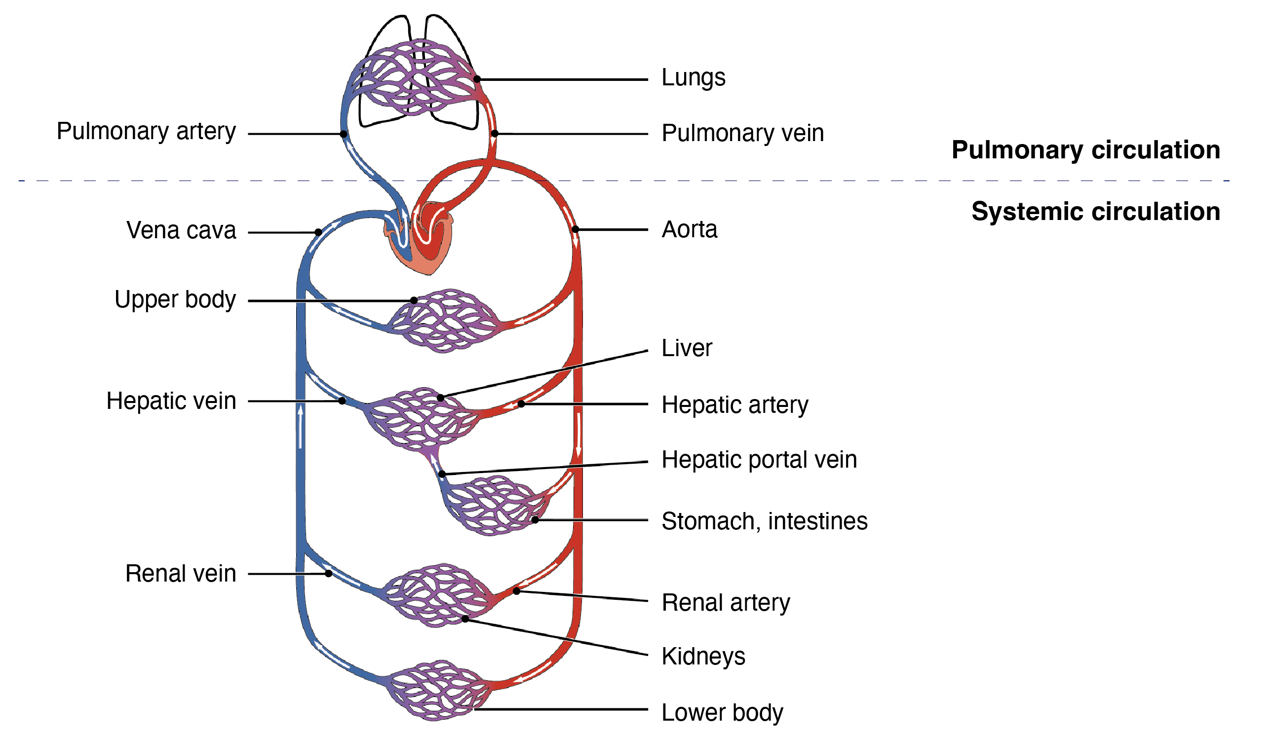
Blood circulation
Gas exchange occurs at the capillary beds, which are networks of tiny blood vessels where oxygen is delivered to cells and carbon dioxide is picked up for removal. These capillary beds are most dense around major organs, like the heart, brain, liver and kidneys.
Pulmonary blood vessels work in the opposite way than the rest of the blood vessels in our bodies. The pulmonary artery carries oxygen-depleted blood, while the pulmonary veins carry oxygen-rich blood.
The atria receives blood and the ventricles pump it out.
The rhythmic thumping of your heart is made by the valves in your heart opening and closing.
Blood in the circulatory system is kept moving in one direction by valves. The heart's natural pacemaker, known as the sinoatrial node regulates the heartbeat, coordinating contractions of the heart muscles to keep a steady rhythm.
Watch this video to learn more about the heart and see it in action.
Now, let's follow the path of the blood through the heart. The superior vena cava receives blood from the head, neck, upper limbs and chest. Meanwhile, the inferior vena cava receives blood from the trunk, viscera and lower limbs. Both the superior and inferior vena cava end up in the right atrium, one of the four chambers of the heart.
The heart not only has four chambers, it also has four valves. The purpose of the valves is to keep blood moving in the right direction and not flow backwards.
Blood exits the right atrium through the tricuspid valve, so called because it has three flaps, and enters the right ventricle. The blood exits the right ventricle through the pulmonary valve and enters the right pulmonary artery.
Again, it is an artery because blood is flowing away from the heart, but it is blue because it lacks oxygen.
The pulmonary artery then splits into the left and right pulmonary arteries, which go to each respective lung. In the lungs, gas exchange occurs. The blood discards carbon dioxide and picks up oxygen.
Now, blood comes back from the lungs through the pulmonary veins, entering the left atrium. Next, the blood is pumped into the left ventricle through the mitral, or bicuspid valve.
Finally, the oxygenated blood leaves the left ventricle through the aortic semi-lunar valuve, entering the aortic arch.
The aorta, which is the largest of all the arteries, distributes the oxygenated blood to the rest of the body. The aortic arch has three major branches, which supply the head and arms with blood.
Then the aorta curls downward behind the heart, forming the descending aorta, which descends through the chest and continues down through the abdomen. In the abdomen, the descending aorta splits to the supply the pelvis and legs with blood.
The components of the circulatory system and their functions are outlined in the table.
| Component | Function |
|---|---|
| Blood | Transports oxygen, nutrients and hormones around the body |
| Blood vessels | Carry blood throughout the body |
| Heart | Pumps blood around the body |
See how well you understand the circulatory system with a quick quiz.
Read the scenario and use the information provided to answer the questions in the quiz.
Zainab is a Year 11 student studying VCE Physical Education. Her class is completing a practical looking at how different types of exercise affect heart rate.
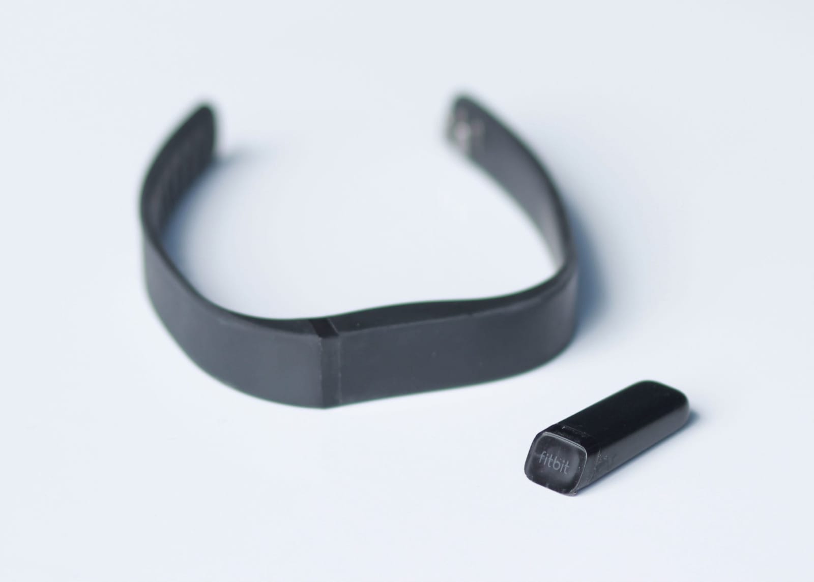
In groups of four, the students take turns completing 10 minutes of jogging, cycling or skipping rope, with 20 minutes of rest in between exercises. They record their resting heart rate and heart rate after 10 minutes of exercise in the table. To measure heart rate, they use a wristband.
| Participant | Resting heart rate (bpm) | Heart rate after 10 min of jogging (bpm) | Heart rate after 10 min of cycling (bpm) | Heart rate after 10 min of skipping (bpm) |
|---|---|---|---|---|
| Zainab | \(72\) | \(120\) | \(110\) | \(130\) |
| Lily | \(68\) | \(115\) | \(108\) | \(125\) |
| Chau | \(75\) | \(122\) | \(116\) | \(128\) |
| August | \(70\) | \(118\) | \(118\) | \(126\) |
National Geographic: Heart 101
Want to learn more about the heart and see how it works? Watch this video.
Cardiovascular system
Use this interactive to explore a 3D model of the circulatory system.
Circulatory system Gizmo
Use this interactive to explore the composition of blood from different blood vessels.
RMIT University acknowledges the people of the Woi wurrung and Boon wurrung language groups of the eastern Kulin Nation on whose unceded lands we conduct the business of the University. RMIT University respectfully acknowledges their Ancestors and Elders, past and present. RMIT also acknowledges the Traditional Custodians and their Ancestors of the lands and waters across Australia where we conduct our business - Artwork 'Sentient' by Hollie Johnson, Gunaikurnai and Monero Ngarigo.
More information