The central nervous system controls the body by processing information and sending signals. Understanding how it works helps us see how our bodies respond to changes and interact with the world. Use this resource to explore how the nervous system keeps everything connected and running smoothly. Also learn about bush medicine that has effects on the central nervous system.
The nervous system
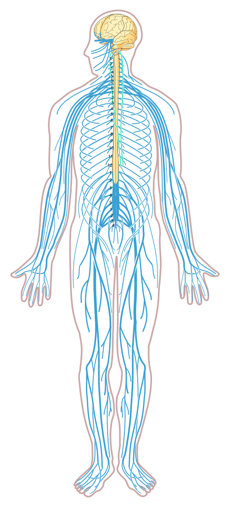
The nervous system is a complex network that controls all the activities in your body. It helps you think, feel, and move by sending messages between your brain and the rest of your body. The nervous system by Medium69 and Jmarchn via Wikimedia Commons licensed under CC BY-SA 4.0.
The nervous system is made up of:
the central nervous system (CNS), which consists of the brain and spinal cord
the peripheral nervous system (PNS), which contains the nerves.
It plays a critical role in helping us interpret information from the environment and coordinate a response.
The central nervous system
The central nervous system (CNS) is the main control centre of the body. It is made up of the brain and spinal cord, which work together to the interpret information we receive from the environment and coordinate a response.
The CNS plays a crucial role in everything from simple reflexes to complex cognitive processes.
The brain
The brain is the most complex organ in the body and serves as the command centre. It manages thoughts, emotions, memory, and decision-making. It also regulates vital functions like breathing and heartbeat.
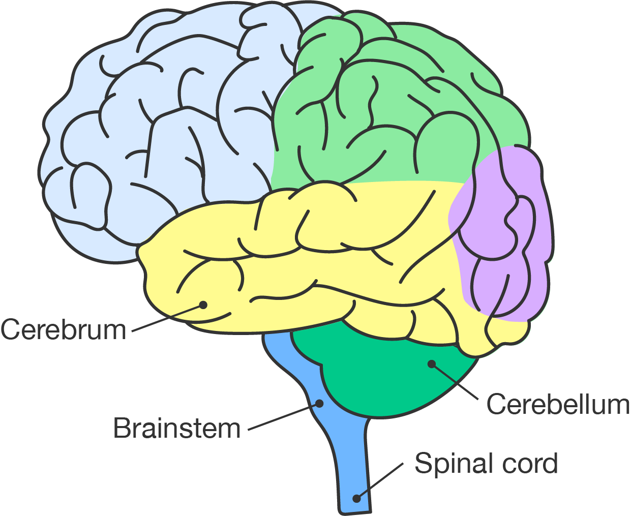
The brain is divided into different regions, each with specialised functions.
Cerebrum (pale blue, pale green, yellow and purple): the largest part of the brain
Cerebellum (green): the section found at the back of the brain, under the cerebrum
Brainstem (blue): the section that connects the brain to the spinal cord
The cerebrum is the largest part of the brain. It is responsible for higher thinking processes and voluntary movement. Divided into two hemispheres (halves), it is organised into lobes that handle different functions like decision-making, sensory information, and regulation of emotions.
Did you know?
The cerebral cortex, the outer layer of the cerebrum, is folded into grooves and ridges to increase its surface area. This allows for more neurones and higher processing power.
The cerebrum allows us to interact with our environment through language, creativity and problem-solving. This forms the basis for intelligence and personality.
The cerebrum is divided into four main lobes. Their functions are explained in the table.
Lobe
Functions
Frontal lobe
Responsible for decision-making, problem-solving, planning and controlling voluntary movements; plays a role in personality and behaviour
Parietal lobe
Processes sensory information related to touch, temperature and pain; helps with spatial awareness and understanding language
Temporal lobe
Involved in hearing, memory and language comprehension; helps process auditory information and is key for recognising faces and objects
Occipital lobe
Primarily responsible for processing visual information; enables us to understand shapes, colours and motion
The lobes work together to make sure that the cerebrum is able to effectively manage complex tasks and responses. Use the 3D model to explore the position of each lobe.
An interactive three-dimensional model of a brain with the lobes of the cerebrum shown in different colours.
Brain model:
Frontal lobe (blue): thinking/imagination
Temporal lobe (green): sound and speech processing
Occipital lobe (red): visual processing
Parietal lobe (yellow)
Pre-frontal lobe (blue): executive and cognitive functions, personality
The cerebellum is found at the back of the brain, under the cerebrum. It focuses on coordination and balance by fine-tuning motor activities (movements that involve our muscles). This allows for smooth and precise movements.
By bringing together sensory inputs from the body and environment, the cerebellum helps us maintain posture and adjust the position of our body during activities. Overall, the cerebellum contributes to effective motor control and learning.
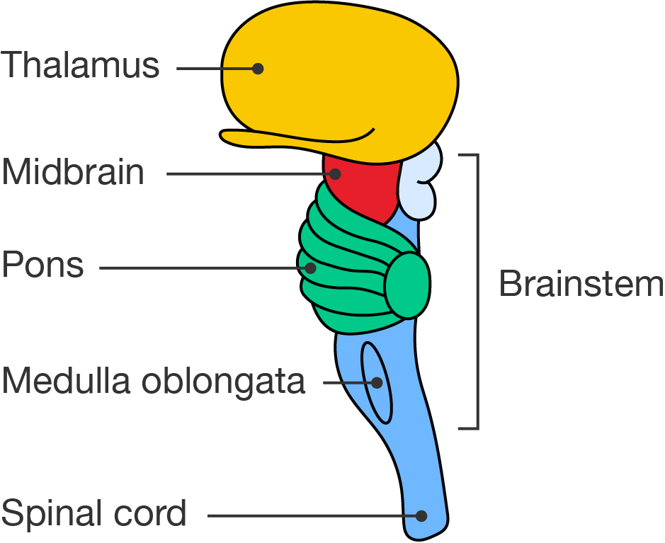
The brainstem connects the brain to the spinal cord. It consists of three parts:
midbrain: controls eye movement and visual and auditory processing; regulates reflexes for seeing and hearing
pons: connects the upper brain regions with the cerebellum; assists in regulating breathing patterns; influence sleep cycles and facial expressions
medulla oblongata: manages vital functions like heartbeat and breathing; coordinates swallowing, sneezing and digestion; serves as a pathway for nerve signals between the brain and body.
Midbrain (red): the section of the brainstem just below the thalamus (yellow)
Pons (green) the section of the brainstem that connects the thalamus and midbrain with the cerebellum
Medulla oblongata (blue): the base of the brainstem, where the brain connects to the spinal cord
The thalamus, sitting above the midbrain, and the spinal cord, positioned directly below the medulla oblongata, are also labeled.
The brainstem also controls reflexes and acts as a pathway for messages between the brain and the body. It helps us survive by managing processes and responses that should be autonomous or reflexive.
The spinal cord
The spinal cord acts as a highway for messages between the brain and the rest of the body. It is responsible for transmitting signals to and from the brain, allowing for fast reflex actions.
The spinal cord is protected by the bones of the vertebral column and consists of nerve fibres that carry impulses to control movements and respond to sensory inputs. Together, the brain and spinal cord form the central nervous system, ensuring that the body operates in a coordinated and efficient manner.
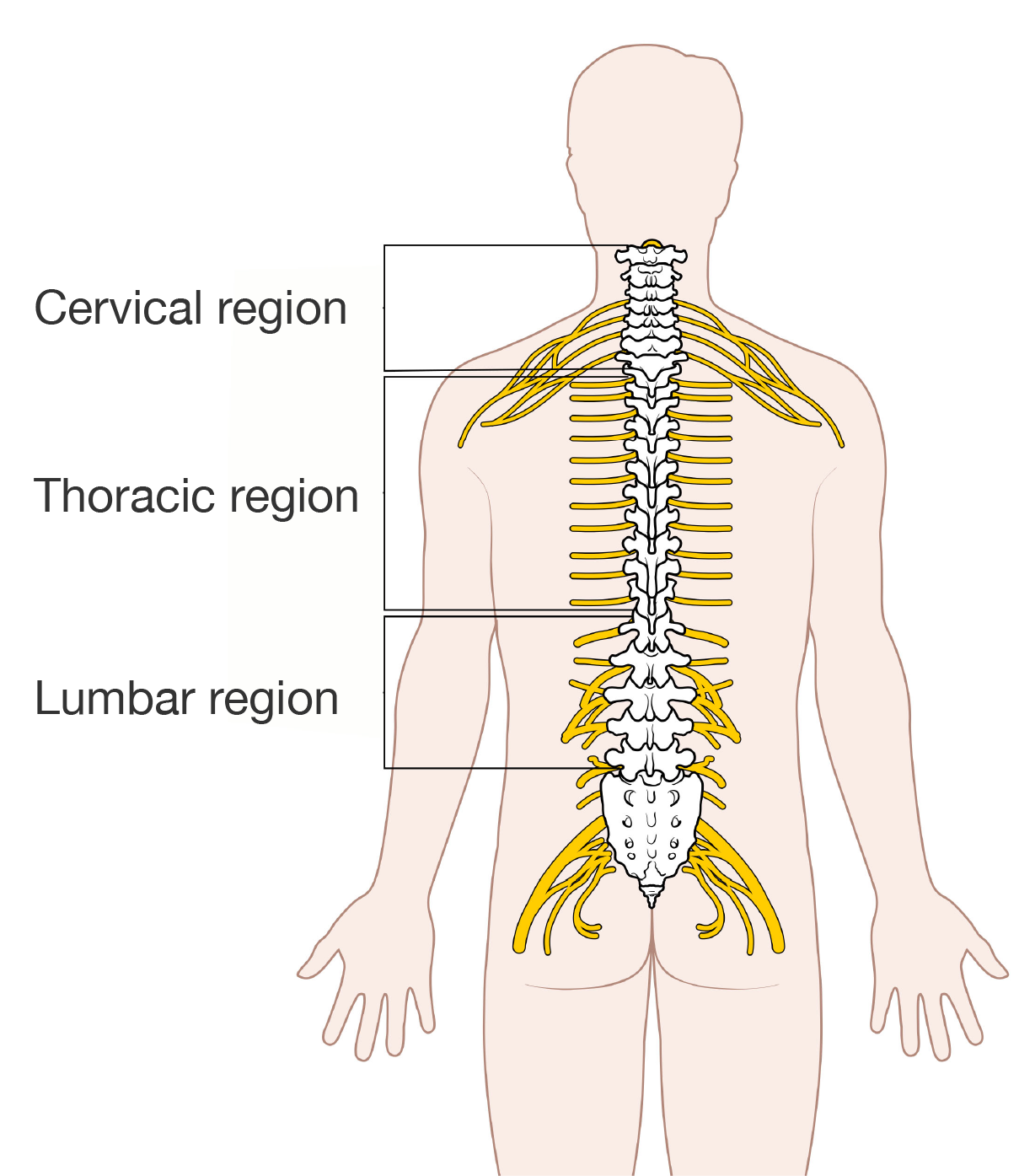
The first 8 segments of the spinal cord below the brain make up the cervical region.
The next 12 segments are the thoracic region.
The following 5 segments are the lumbar region.
Then, there are 5 segments in the sacral region and just 1 coccygeal segment at the bottom of the spinal cord, which are not labeled in the figure.
Did you know?
The spinal cord doesn't always rely on the brain to tell it what to do. Sometimes, it sends signals to the muscles on its own. These signals are called reflexes.
Indigenous knowledges in life science
Corkwood tree as a sedative
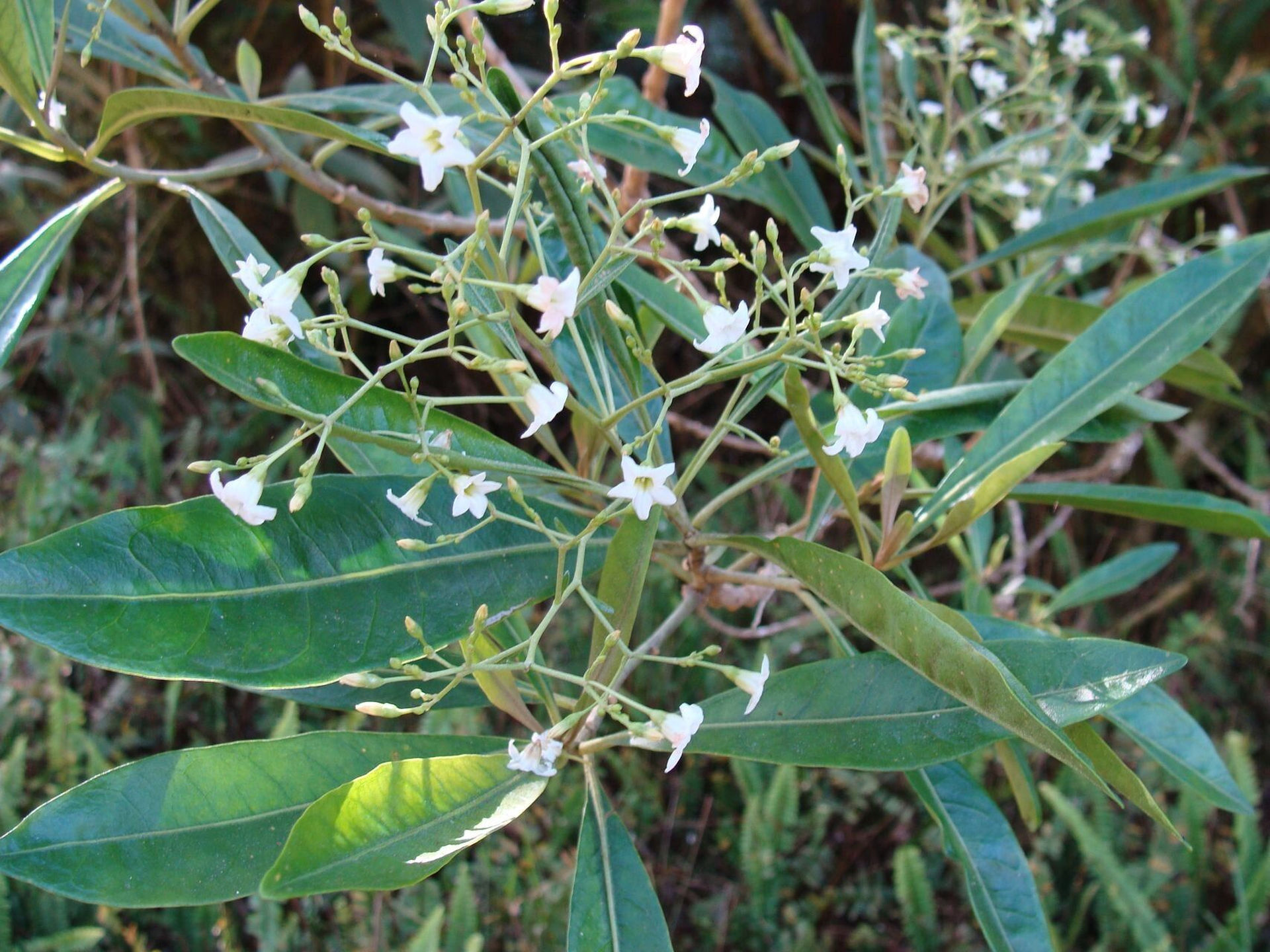
Aboriginal peoples in the Eastern regions of Australia developed a bush medicine from soft corkwood tree. It was used in many ways, such as a remedy for seasickness by fisherman, in ceremony to connect with spiritual realm, and as a sleeping potion.
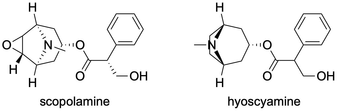
The medicine was adopted by the Allies in WWII to treat seasickness in their soldiers, keeping them unconscious as they sailed across the English Channel. Later, this was used to produce drugs called scopolamine (for motion and seasickness) and hyoscyamine (for abdominal spasms), which are now also very important in eye surgery medicine. These compounds are structurally related and belong to a class of chemicals known as the tropane alkaloids. They are commercially harvested from the leaves of the native soft corkwood tree (Duboisia myoporoides) and its hybrid varieties and this has boomed into a multi-million-dollar industry in Queensland.
Scopolamine and hyoscyamine chemical structures, image by RMIT, licensed under CC BY-NC 4.0
These compounds bind to the active sites of muscarinic receptors, which are present across the central and peripheral nervous system, and block the activity of acetylcholine. They are also muscle relaxants and sedatives.
See how well you understand the components of the central nervous system and their roles with a quick quiz.
Data drill
Read the scenario and use the information provided to answer the questions in the quiz.
In 2014, researchers at the University of Sousse and the University of Sfax collaborated on a project looking at the effect of caffeine ingestion on reaction time after 36 hours of sleep deprivation. Thirteen healthy male physical education students completed four test sessions at 6 pm:
placebo during a baseline night (bedtime from 10:30 pm to 7 am)
caffeine ingestion during a baseline night (bedtime from 10:30 pm to 7 am)
placebo during a night of 36 h of sleep deprivation
caffeine ingestion during a night of 36 h of sleep deprivation.
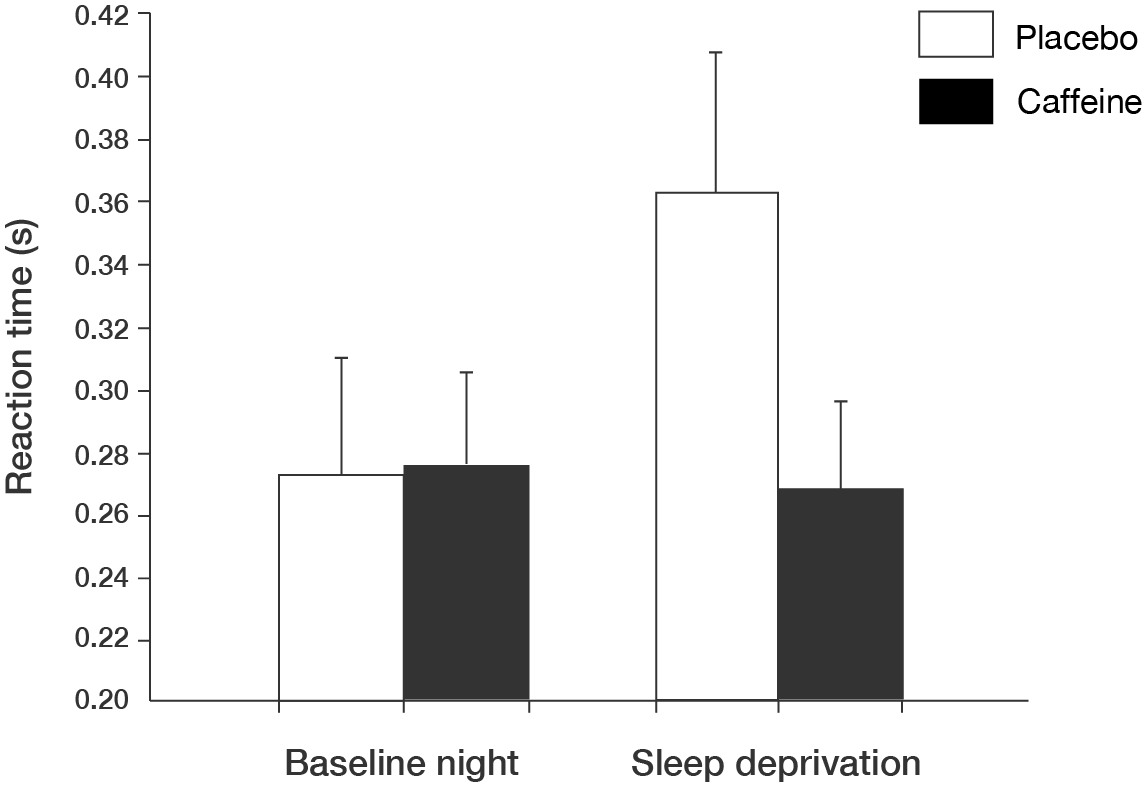
Participants were tested on their performance of serveral physical activites, including a reaction time test. For the reaction time test, they were required to press a key on a microcomputer when a visual stimulus appeared. The results for the test are shown in the figure.
Bar chart showing results of reaction time test
A vertical bar chart measuring reaction time on with a baseline night of sleep or sleep deprivation, with placebo or caffeine ingestion.
All groups are shown on the x-axis.
The first bar is baseline night with placebo.
The second bar is baseline night with caffeine.
The third bar is sleep deprivation with placebo.
The fourth bar is sleep deprivation with caffeine.
The y-axis is labelled "Reaction time (s)", starting at 0.20 seconds and extending to 0.42 seconds, with 0.02 second increments.
The data is shown in the table.
Condition
Reaction time with placebo (mean ± SD)
Reaction time with caffeine (mean ± SD)
Baseline night
\(0.275 \pm 0.040\)
\(0.278 \pm 0.030\)
Sleep deprivation
\(0.362 \pm 0.050\)
\(0.268 \pm 0.030\)
Graph adapted from Souissi M, Chtourou H, Abedelmalek S, Ghozlane IB and Sahnoun (2014), 'The effects of caffeine ingestion on the reaction time and short-term maximal performance after 36h of sleep deprivation', Physiology & Behavior, 131:1-6, doi: 10.1016/j.physbeh.2014.04.012.