The muscular system
Use this 3D interactive to explore the different muscles in the human body.
The muscular system provides support and facilitates movement of the body. Understanding how it works helps us see how the body performs physical activities and stays stable and strong. Use this resource to explore the components and roles of the muscular system.
The muscular system consists of all of the muscules in your body and the tissues that support them. It works with the skeletal system to help you move, maintain your posture and produce heat.
It also protects the internal organs and assist in critical functions like breathing and digestion.
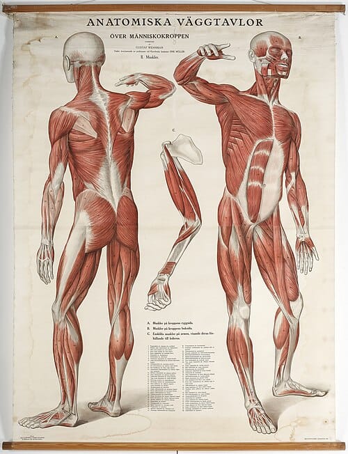
Skeletal muscle is a type of muscle that is attached to bones. It allows us to move voluntarily, like walk and lift things. It also helps us maintain our posture and generates heat to keep us warm.
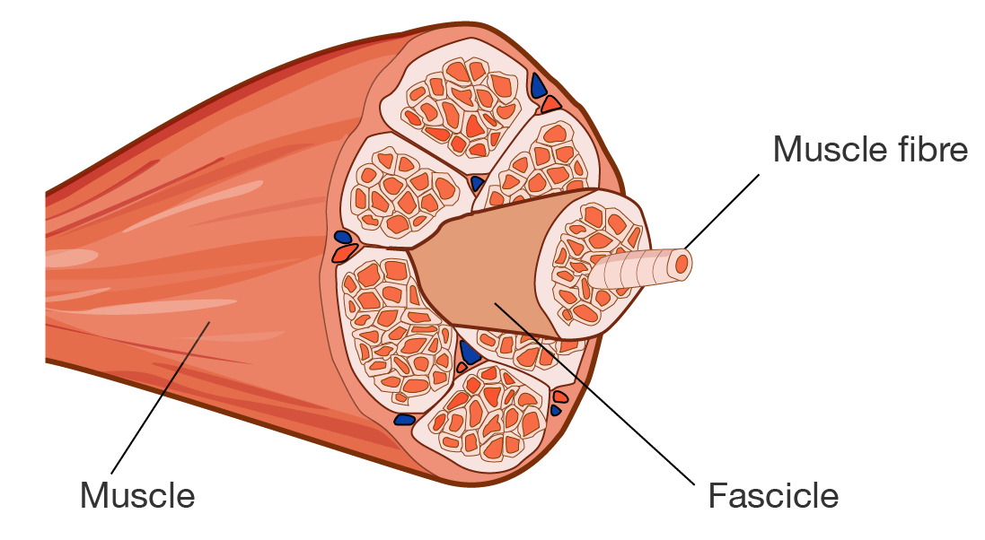
Skeletal muscle is composed of fibres bundled together by connective tissues. It is striated, meaning that it is made of parallel light and dark bands. Rod-like microfibrils also run parallel to the muscle fibre. These are responsible for muscle contraction.
A diagram of a cross-section of skeletal muscle showing its structure. Each muscle fibre is bundled together to form a fascicle. Each fascicle is then bundled up to form the muscle.
Skeletal muscle
The eye muscles are the fastest muscles in the body. They can contract in less than \(\dfrac{1}{100}\)th of a second, allowing you to blink over \(20,000\) times a day.
The contraction of muscles is controlled by the nervous system. The brain coordinates the motor neurones, which send signals to the skeletal muscle to contract. Muscles work in pairs, where one muscle contracts while the other relaxes to coordinate movement.
Examples of skeletal muscle include the triceps, which are at the back of your arm and the gluteus maximus, also known as the glutes or butt, which is the largest muscle in the body. You can go to this link to see a list of the skeletal muscles in the human body.
To make our skeletal muscle stronger, we need to exercise regularly. This improves flexibility and endurance.
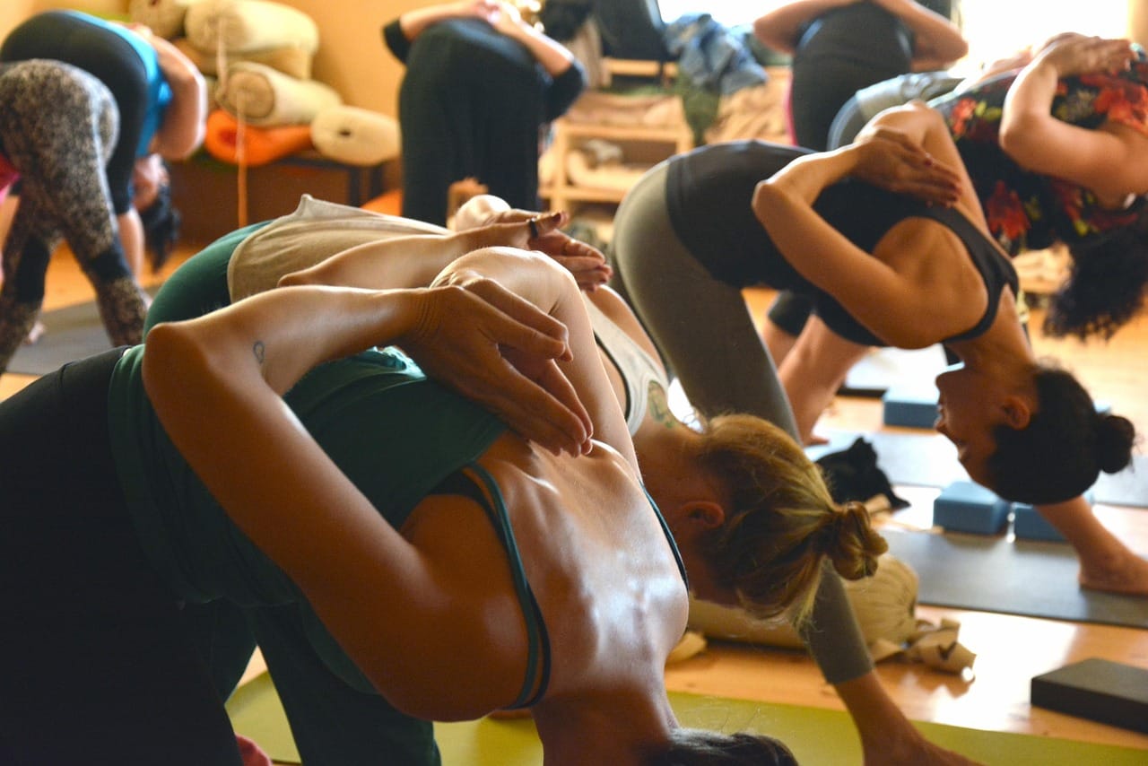
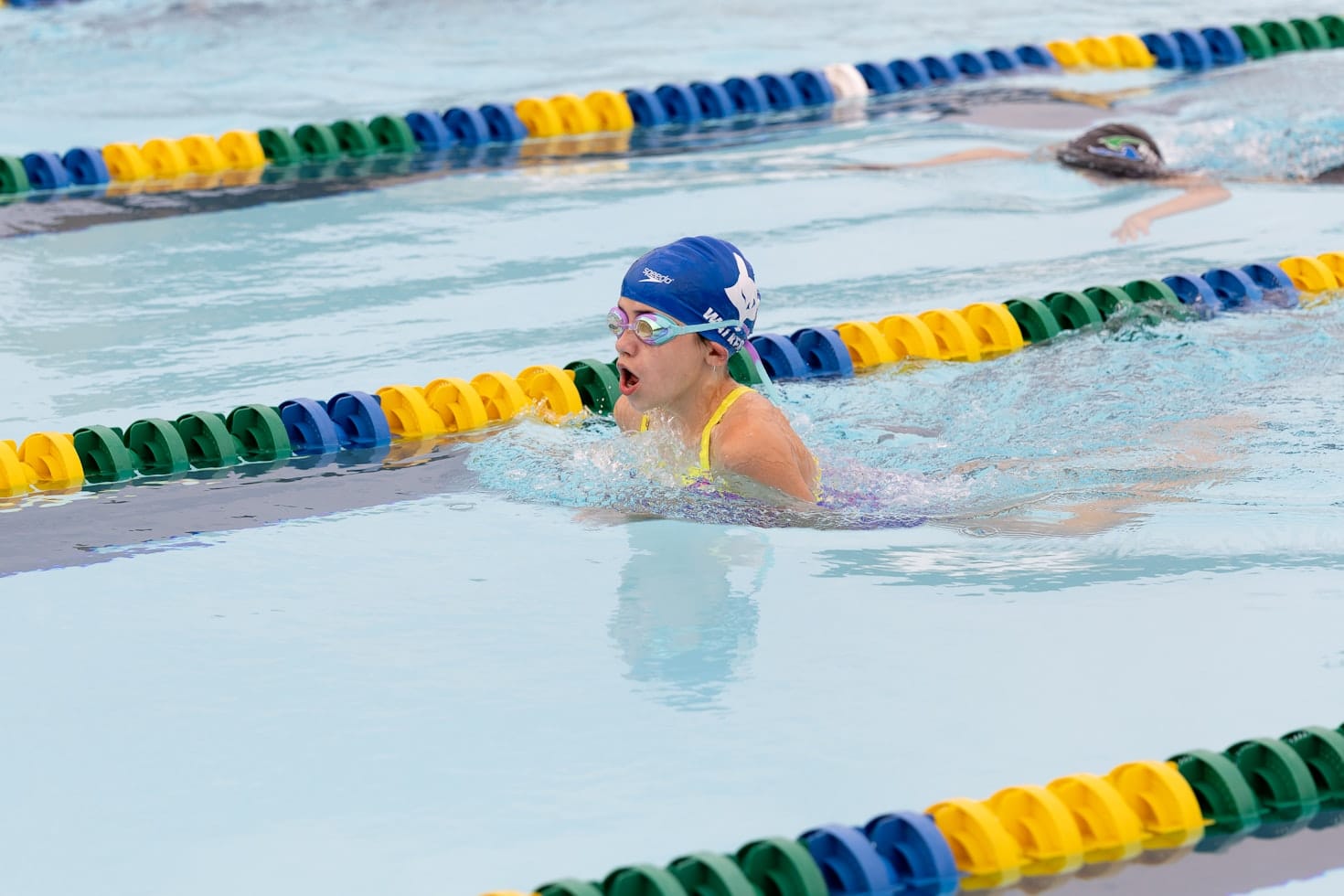
Yoga class by shushipu via Pixabay; Swimmer by Johanna Steppan via Unsplash
Our skeletal muscles use adenosine triphospate (ATP) for energy during contraction. Learn more about ATP and cellular respiration on this page.
The body can generate this energy through two types of metabolism:
The process of ATP generation also produces heat, which the body can use to regulate its temperature.
Tendons connect bones to skeletal muscle. They are mostly made up of collagen fibres, which make them tough and flexible.
Tendons work like cords, allowing muscles to pull bones effectively. They help transfer force and stabilise joints during movement.
Smooth muscle is found in the walls of organs like the stomach, intestines and blood vessels. Unlike skeletal muscle, it moves on its own, meaning that we don't control it consciously.
Smooth muscle contracts slowly to move substances through the digestive tract and regulate blood flow. It is particularly important for digestion and blood pressure regulation because it can stay contracted over longer periods of time. Smooth muscle can do this because it uses less ATP.
Smooth muscle in your digestive tract helps move food along through a process called peristalsis. This muscle contraction can occur even when you're upside down, which is why you can still swallow and digest food in zero gravity.
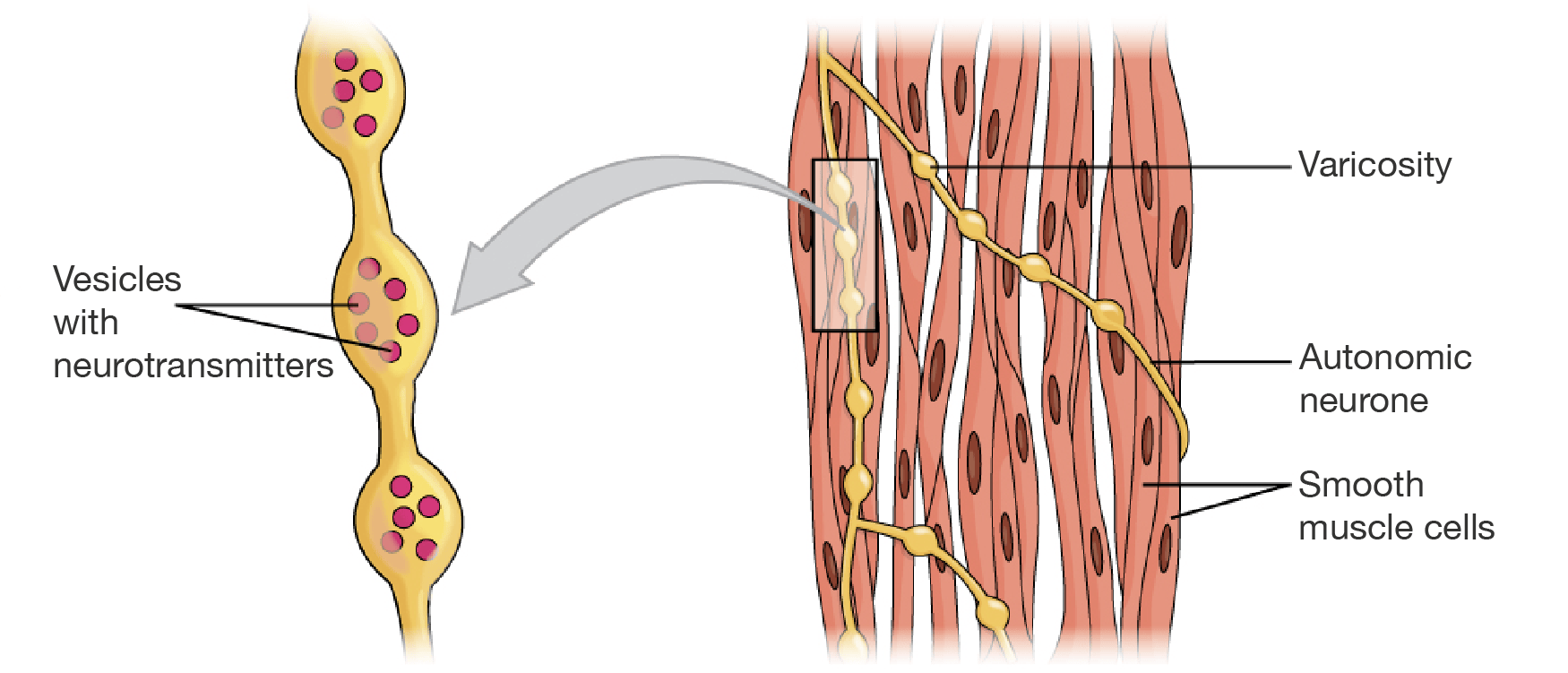
While skeletal muscles are made up of bundles of fibres that work together, smooth muscle is made up of individual cells that contract as one tissue in organs. Because of this, smooth muscle looks more uniform.
A diagram showing the organisation of smooth muscle, with a zoomed in section of neurones. On the right is a section of smooth muscle, as shown as spindle-shaped cells. They are not arranges in tubes like skeletal muscle. The cells are labelled "Smooth muscle cells". Wrapped around the cells are thin structures labelled "Autonomic neurone". These neurones have sections where they bulge and bulbed areas are labelled "Varicosity". A section of the autonomic neurone with varicosity is highlighted with a rectangle. There is an arrow pointing from this rectangle to the image on the left. The image on the left is a zoomed in section of the varicosity. Within the bulbed regions are small balls with the label "Vesicles with neurotransmitters".
Smooth muscle
The smooth muscle is wrapped in neurons, meaning that it partly relies on the nervous system to coordinate its action. As well as this, it responds to hormones. For example, the hormone gastrin stimulates the contraction of smooth muscle in the stomach to help churn food.
Cardiac muscle is found in the walls of the heart and is responsible for its rhythmic contractions. Like smooth muscle, it works involuntarily. It beats tirelessly, pumping blood throughout the body and is crucial for keeping our organs supplied with oxygen and nutrients.
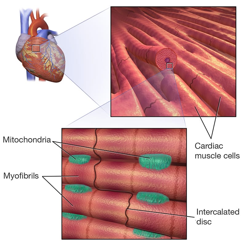
Cardiac muscle has a unique structure. It is striated like skeletal muscle but forms interconnected networks called intercalated disks. This allows rapid transmission of electrical signals for the heart to contract efficiently and uniformly. They also have a high density of mitochondria to support constant contraction. Cardiac muscle is regulared by the sinoatrial node.
A diagram showing the organisation of cardiac muscle. There are three images. The first image is of a whole heart. There is a small rectangle drawn on the surface of the heart, corresponding to the second image. The second image is a zoomed up look at the heart. It shows the "Cardiac muscle cells" labelled. They are connected in a network, with thick black lines separating some strands of muscle. One strand of cardiac muscle is cut to reveal its cross-section. A rectangle is drawn at the cross-section, corresponding to the third image. The third image is a zoomed up look at the cardiac muscle fibre. It shows four fibres lined up, each labelled "Myofibrils". A thick black line runs across the fibres, labelled "Intercalated disc". Between the myofibrils are green blob-like structures, labelled "Mitochondria".
Cardiac muscle
The components of the muscular system and their functions are outlined in the table.
| Component | Function |
|---|---|
| Skeletal muscle | Attached to bones; enables voluntary movements like walking and lifting |
| Tendons | Connects muscles to bones; transfers force to move bones, stabilising joints |
| Smooth muscle | Found in organ walls; controls involuntary movements like digestion and blood flow |
| Cardiac muscle | Located in the heart; pumps blood continuously without conscious control |
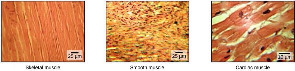
Three microscopy images showing skeletal muscle, smooth muscle and cardiac muscle. Each image has a horizontal bar to indicate the scale. The width of the bar is 25 micron.
See how well you understand the components and functions of the muscular system with a quick quiz.
Read the scenario and use the information provided to answer the questions in the quiz.
A group of researchers in Japan conducted a meta-analysis of randomised controlled trials to study the relationship between total protein intake and muscle strength, with and without resistance training.
Their results are shown in the graphs. The solid lines represent the averages and the dashed lines represent the 95% confidence interval.
On the left is a line graph titled "With resistance training". There are three data lines on the graph, a solid line labelled "Average" and two dashed lines labelled "95% confidence interval". There is another line at x equals 0 to show where baseline is. The x-axis is labelled "Total protein intake (grams per kg of bodyweight per day)", starting at 0.5 g and going all the way to 4, with increments of 0.5. The y-axis is labelled "Change in muscle strength (%)", starting at negative 20 and going all the way to 30, with increments of 5. In the graph, the "Average" line starts at around 0.7 grams per kg of bodyweight per day which gives a 19.5% change in muscle strength. As total protein intake increases, the change in muscle strength increases until a change of 24.5% with 1.5 grams per kg of bodyweight per day. From there, the change in muscle strength gradually decreases until it reaches 15.0% with 3.8 grams per kg of bodyweight per day. The 95% confidence interval lines show the spread of data around the mean. As we approach the peak muscle strength, the data becomes less spread. Then, as the total protein intake increases further, the data becomes more spread. At the maximum protein intake of 3.8 grams per kg of bodyweight per day, the 95% confidence interval is between 10% and 20%. On the right is a line graph titled "Without resistance training". The format is the same as the first graph, but with different data. In the graph, the "Average" line starts at around 0.7 grams per kg of bodyweight per day which gives a 4% change in muscle strength. As total protein intake increases, the change in muscle strength increases until a change of 7% with 1.3 grams per kg of bodyweight per day. Then, the change in muscle strength gradually decreases until it reaches 3% at 2 grams per kg of bodyweight per day. The 95% confidence interval decreases until the peak change in muscle strength is reached, then increases again. The spread of data at 0.7 and 2.0 grams per kg of bodyweight per day is similar. The data is shown in the table.
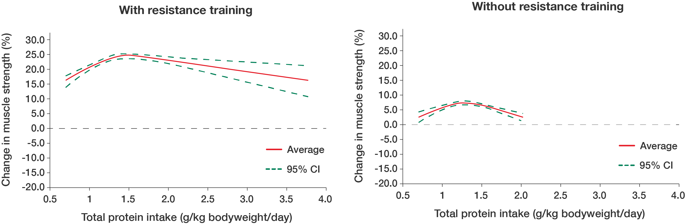
Line graph showing change in muscle strength with resistance training
Line graph showing change in muscle strength with no resistance training
Total protein intake (g/kg bodyweight/day)
With resistance training (Change in muscle strength %)
Without resistance training (Change in muscle strength %)
\(0.7\)
\(19.5\)
\(4.0\)
\(1.0\)
\(22.0\)
\(6.0\)
\(1.5\)
\(24.5\)
\(7.5\)
\(2.0\)
\(23.0\)
\(3.0\)
\(2.5\)
\(20.0\)
Not tested
\(3.0\)
\(18.0\)
Not tested
\(3.5\)
\(16.0\)
Not tested
\(3.8\)
\(15.0\)
Not tested
Graphs adapted from Tagawa R, Watanabe D, Ito K, Otsuyama T, Nakayama K, Sanbongi C and Miyachi M (2022), 'Synergistic Effect of Increased Total Protein Intake and Strength Training on Muscle Strength: A Dose-Response Meta-analysis of Randomized Controlled Trials', Sports Medicine – Open, 8(110), doi: 10.1186/s40798-022-00508-w, licensed under CC BY 4.0
Images on this page by RMIT, licensed under CC BY-NC 4.0

RMIT University acknowledges the people of the Woi wurrung and Boon wurrung language groups of the eastern Kulin Nation on whose unceded lands we conduct the business of the University. RMIT University respectfully acknowledges their Ancestors and Elders, past and present. RMIT also acknowledges the Traditional Custodians and their Ancestors of the lands and waters across Australia where we conduct our business - Artwork 'Sentient' by Hollie Johnson, Gunaikurnai and Monero Ngarigo.
More information