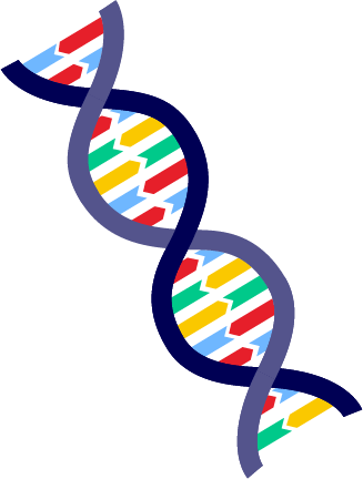
DNA carries the genetic instructions needed for the growth, development and functioning of all living organisms. You must understand the structure of DNA to grasp how genetic information is passed down and used in biological processes. This is crucial in fields like medicine, where it aids in developing treatments and in biotechnology, where it drives innovation. Use this resource to learn about the structure of DNA.
DNA is composed of two long strands of nucleotides twisted around each other. Go back to Macromolecules if you need to review nucleic acids and nucleotides.
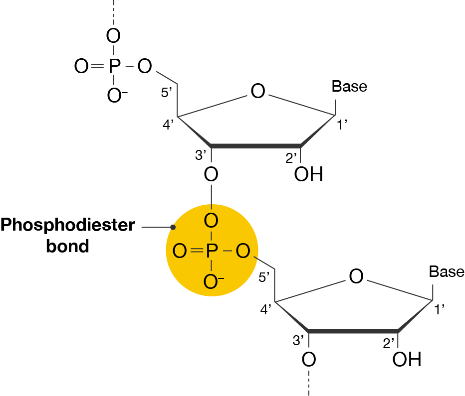
The nucleotides in a strand of DNA are connected by phosphodiester bonds between the third carbon (3’ or three prime) of a sugar of one nucleotide and the phosphate attached to the fifth carbon (5’ or five prime) of the sugar on the next nucleotide. Nucleotides are only added to the 3’ end, so the direction of DNA is 5’ to 3’. DNA replication and transcription also occur in this direction.
Phosphodiester bond
A section of a nucleic acid strand is shown, consisting of two nucleotides. The phosphodiester bond highlighted. This is a phosphate group that connects the two nucleotides.
The phosphate group forms a bridge between the 3 prime carbon of the top sugar and the 5 prime sugar of the bottom sugar. This group is made up of a central phosphorus atom bonded to four oxygen atoms: one double-bonded oxygen (O=P), two single-bonded oxygens linking to the sugars (O-P-O) and one negatively charged oxygen (O-).
Specific sequences of nucleotides are called genes, which code for specific proteins. You will learn about how DNA is used to make proteins in Protein synthesis.
The double helix
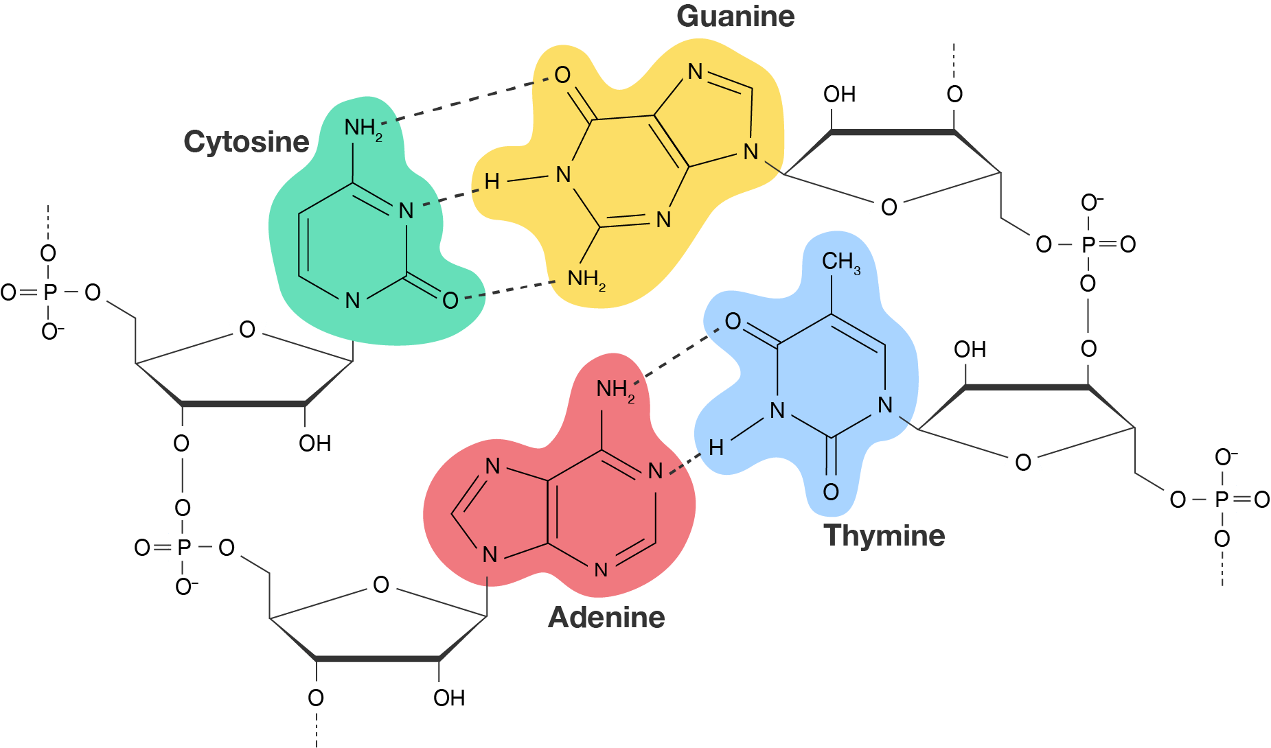
DNA has a double helix structure. This is formed through complementary base pairing. Adenine (A) pairs specifically with thymine (T), and cytosine (C) pairs specifically with guanine (G). DNA is often referred to as a twisted ladder as these bases form hydrogen bonds with each other to create "rungs". The two strands run in opposite directions causing the sugar-phosphate backbone to alternate.
Guanine and cytosine pair via three hydrogen bonds, whereas there are only two hydrogen bonds between adenine and thymine. The stronger association between guanine and cytosine gives DNA its twisted structure.
Complementary base pairing
A segment of double-stranded DNA, with each strand containing two nucleotides. The left strand has a cytosine nucleotide and an adenine nucleotide. The right strand has a guanine nucleotide and a thymine nucleotide.
The cytosine nucleotide (highlighted in green) pairs with a guanine nucleotide (highlighted in yellow) through three hydrogen bonds, shown in dashed lines. The adenine nucleotide (highlighting in red) pairs with a thymine nucleotide (highlighted in blue) through two hydrogen bonds, also shown as dashed lines.
You can use this interactive to see a 3D model of the DNA structure.
A three-dimensional model of a segment of DNA with atoms shown as coloured sticks.
DNA double helix model
Phosphate is shown is gold, oxygen is red, nitrogen is blue, carbon is teal and hydrogen is white. The atoms are arranged to form two strands of nucleotides. The strands form a twisted ladder structure called the double helix.
Chromosomes
In the nucleus, the DNA is coiled up into a condensed structure which protects the DNA during cell division.
Packaging proteins called histones bind to the DNA molecule. When a section of DNA is wrapped around a cluster of eight histones, it is called a nucleosome. Chains of nucleosomes are further coiled into a spiral called a solenoid. The solenoids coil to form the fibres of chromatin.
When the cell is not dividing, DNA is present in the nucleus as chromatin. When it is ready to divide, it further condenses to form chromosomes.
A three-dimensional model of DNA structure, showing the different levels of organisation.
DNA structure model
Strands of DNA are organised into a double helix structure.
The double helix is coiled around clusters of four histone proteins, shown in pink and yellow.
The histones are further coiled to form solenoids.
Solenoids are coiled even further to eventually form chromatin.
Chromatin condenses into chromosomes.
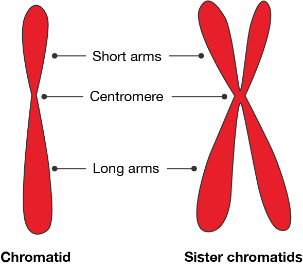
A chromosome consists of a chromatid, which has a short arm and a long arm. Between the arms is a constricted region called a centromere. One chromosome is one DNA molecule.
When DNA is being replicated, the chromosome duplicates and consists of two sister chromatids joined at the centromere.
Chromatids
A simplified drawing of chromatids. A single chromatid is shown on the left. It has a pinched region in the middle, labelled the "Centromere". The centromere is slightly higher on the chromatid, so the top section is shorter than the bottom. These correspond to the "Short arm" and "Long arm", respectively.
On the right, sister chromatids are shown. It consists of two chromatids joined at the constricted region. The centromere, short arms and long arms are also labelled, matching the drawing on the left.
Exercise
See how well you understand the structure of DNA with a quick quiz.
Ready for some more? Practise your skills by determining the sequence of the DNA strand that is complementary to the following strands:
TAC–CGA–CCA–ATT–TAG–TTA–CCC
CCG–AAG–CTG–ATG–GGA–CAG–ACG
GTT–AGA–CTA–TAA–GCG–ACT–TTT
AAT–ACC–CAG–TTG–CGG–AAA–TCA
AGA–CTC–TGA–CGG–TAC–GAA–CTG
TCT–GAG–ACT–GCC–ATG–CTT–GAC
TGC–AAG–TTC–GGA–CCA–ATG–TAC
ATG–GCT–GGT–TAA–ATC–AAT–GGG
GGC–TTC–GAC–TAC–CCT–GTC–TGC
CAA–TCT–GAT–ATT–CGC–TGA–AAA
TTA–TGG–GTC–AAC–GCC–TTT–AGT
TCT–GAG–ACT–GCC–ATG–CTT–GAC
GCT–ATG–CCA–TTG–AAT–GGC–CTA
ACG–TTC–AAG–CCT–GGT–TAC–ATG
Data drill
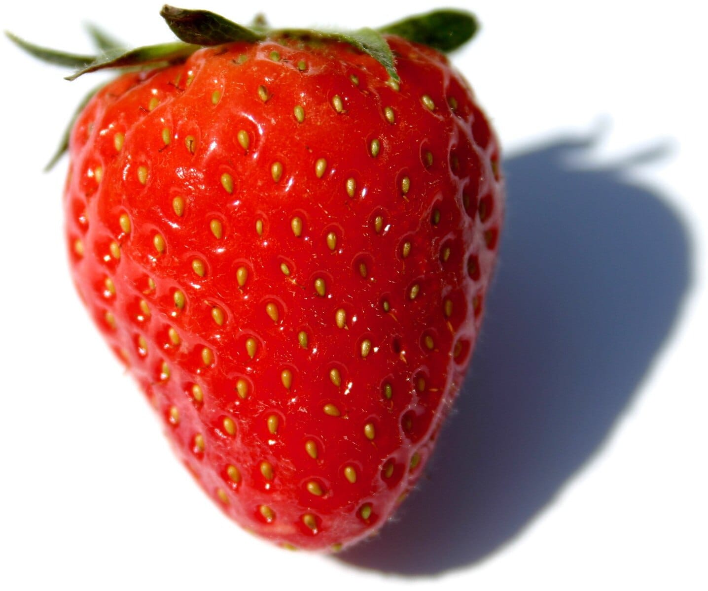
Read the scenario and use the information provided to answer the questions in the quiz. Strawberry, by FoeNyx via Wikimedia Commons, licensed under CC BY-SA 2.5
A class of biology students have recently completed a practical where they extracted DNA from strawberries. Each group of students used half a strawberry each, completed the extraction and measured the concentration of DNA using a spectrophotometer.
The class data is collated in the following table.
